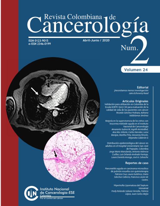
Lipomatous hypertrophy of the interatrial septum. A case report
DOI:
https://doi.org/10.35509/01239015.347Keywords:
Pathology, Diagnosis, Heart Tumors., Hypertrophy, atrial septumAbstract
Lipomatous Hypertrophy of the Interatrial Septum (LHIS) is a rare and benign cardiac entity that is characterized by the accumulation of adipose tissue within some segments of the interatrial septum. Patients are generally asymptomatic, and these lesions are discovered incidentally by imaging studies performed for other reasons, or in the context of an autopsy. In these patients, there have been described cases of sudden death due to disturbance of the heart rhythm. The differential diagnosis of LHIS mainly includes cardiac tumors. Here we present a case of a 61-year-old patient in whom, after a cardiac magnetic resonance study performed for an abnormal heart rhythm, it was documented a mass in the atrial septum. The patient was taken to surgery, and the histopathological study of the lesion confirmed the diagnosis. We conduct a review of the clinical and pathological characteristics of LHIS.
References
Prior JT. Lipomatous hypertrophy of cardiac interatrial septum. Arch Pathol. 1964;78:11-5
Nadra I, Dawson D, Schmitz SA, Punjabi PP, Nihoyannopoulos P. Lipomatous hypertrophy of the interatrial septum: a commonly misdiagnosed mass often leading to unnecessary cardiac surgery. Heart. 2004;90:e66. https://doi.org/10.1136/hrt.2004.045930
Hejna P, Janík M. Lipomatous hypertrophy of the interatrial septum: a possibly neglected cause of sudden cardiac death. Forensic Sci Med Pathol. 2014;10:119-21. https://doi.org/10.1007/s12024-013-9480-0
Miraglia E, Visconti B, Bianchini D, Calvieri S, Giustini S. An uncommon association between lipomatous hypertrophy of the interatrial septum (LHIS) and Dercum’s disease. Eur J Dermatol. 2013;23:406-7. https://doi.org/10.1684/ejd.2013.2016
O’Connor S, Recavarren R, Nichols L, Parwani AV. Lipomatous hypertrophy of the interatrial septum: an overview. Arch Pathol Lab Med. 2006;130:397-9.
Heyer CM, Kagel T, Lemburg SP, Bauer TT, Nicolas V. Lipomatous hypertrophy of the interatrial septum: a prospective study of incidence imaging findings, and clinical symptoms. Chest. 2003;124:2068-73. https://doi.org/10.1378/chest.124.6.2068
Xanthos T, Giannakopoulos N, Papadimitriou L. Lipomatous hypertrophy of the interatrial septum: a pathological and clinical approach. Int J Cardiol. 2007;121:4-8. https://doi.org/10.1016/j.ijcard.2006.11.150
Tutar E, Çiftçi O, Fitoz S, Kendirli T, Ödek Ç, Uçar T, et al. Lipomatous hypertrophy of the interatrial septum in a child with atrial tachycardia. Pediatr Int. 2016;58:523-5. https://doi.org/10.1111/ped.12917
Laura DM, Donnino R, Kim EE, Benenstein R, Freedberg RS, Saric M. Lipomatous atrial septal hypertrophy: a review of its anatomy, pathophysiology, multimodality imaging, and relevance to percutaneous interventions. J Am Soc Echocardiogr. 2016;29:717-23. https://doi.org/10.1016/j.echo.2016.04.014
Escobar C, Jaramillo M, Tenorio L, Molina C, Saldarriaga M, Arango A. Ecocardiografía transesofágica en el estudio de pacientes con eventos cerebrovasculares en quienes se sospecha origen cardiovascular embólico. Rev Col Cardiol. 2003;10:199-204.
Miller DV, Tazelaar HD. Cardiovascular pseudoneoplasms. Arch Pathol Lab Med. 2010;134:362-8.
Bois M, Bois JP, Anavekar NS, Oliveira AM, Maleszewski JJ. Benign lipomatous masses of the heart: a comprehensive series of 47 cases with cytogenetic evaluation. Hum Pathol. 2014;45:1859-65. https://doi.org/10.1016/j.humpath.2014.05.003
Rob D, Kuchynka P, Palecek T, Cerny V, Masek M, Vitkova I, et al. A rare case of regressively changed lipomatous hypertrophy of the interatrial septum presenting with anemia and recurrent fever. Cardiovasc Pathol. 2016;25:161-4. https://doi.org/10.1016/j.carpath.2015.09.002
Calé R, Andrade MJ, Canada M, Hernandez ER, Lima S, Abecasis M, et al. Lipomatous hypertrophy of the interatrial septum: report of two cases where histological examination and surgical intervention were unavoidable. Eur J Echocardiogr. 2009;10:876-9. https://doi.org/10.1093/ejechocard/jep080
Bielicki G, Lukaszewski M, Kosiorowska K, Jakubaszko J, Nowicki R, Jasinski M. Lipomatous hypertrophy of the atrial septum - a benign heart anomaly causing unexpected surgical problems: a case report. BMC Cardiovasc Disord. 2018;18:152. https://doi.org/10.1186/s12872-018-0892-3
How to Cite
Downloads
Downloads
Published
Issue
Section
License
Todos los derechos reservados.


