Image fusion and hippocampus protection in radiotherapy treatment of brain metastases
Keywords:
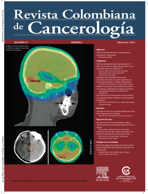
Tomography, X-ray computed, Magnetic resonance imaging, Image superimposition, Radiosurgery, Brain metastases, HippocampusAbstract
CT simulation images are the current standard in external beam radiotherapy planning systems. However, the limitations of images obtained from CT scanning include their low contrast and low specificity in the identification and characterization of tumor lesions and some central nervous system structures. The new algorithms implemented in radiotherapy planning systems allow image fusion to be performed using MRI images and CT simulation images. It also allows structures like the hippocampus to be defined and protected, by administering lower doses to this area and higher doses to the tumor volume, thus decreasing side effects arising from whole brain radiotherapy treatment. Images corresponding to this treatment technique are presented in this article.
Author Biographies
Elizabeth Alwers, Centro de Control de Cáncer Ltda
Centro de Control de Cáncer Ltda., Bogotá D. C., Colombia
Iván Bobadilla, Centro de Control de Cáncer
Centro de Control de Cáncer Ltda., Bogotá D. C., Colombia
Jaider Vásquez, Centro de Control de Cáncer
Centro de Control de Cáncer Ltda., Bogotá D. C., Colombia
Javier Cifuentes, Centro de Control de Cáncer
Centro de Control de Cáncer Ltda., Bogotá D. C., Colombia
Felipe Torres, Centro de Control de Cáncer
Centro de Control de Cáncer Ltda., Bogotá D. C., Colombia
References
Nayak L, Lee EQ, Wen PY. Epidemiology of brain metastases. Curr Oncol Rep. 2012;14(1):48-54.
https://doi.org/10.1007/s11912-011-0203-y
Gállego Pérez-Larraya J, Hildebrand J. Brain metastases. Handb Clin Neurol. 2014;121:1143-57.
https://doi.org/10.1016/B978-0-7020-4088-7.00077-8
Kondziolka D, Patel A, Lunsford LD, Kassam A, Flickinger JC. Stereotactic radiosurgery plus whole brain radiotherapy versus radiotherapy alone for patients with multiple brain metastases. Int J Radiat Oncol. 1999;45(2):427-34.
https://doi.org/10.1016/S0360-3016(99)00198-4
Andrews DW, Scott CB, Sperduto PW, Flanders AE, Gaspar LE, Schell MC, et al. Whole brain radiation therapy with or without stereotactic radiosurgery boost for patients with one to three brain metastases: phase III results of the RTOG 9508 randomised trial. Lancet. 2004;363(9422):1665-72.
https://doi.org/10.1016/S0140-6736(04)16250-8
Mehta MP, Tsao MN, Whelan TJ, Morris DE, Hayman JA, Flickinger JC, et al. The American Society for Therapeutic Radiology and Oncology (ASTRO) evidence-based review of the role of radiosurgery for brain metastases. Int J Radiat Oncol Biol Phys. 2005;63(1):37-46.
https://doi.org/10.1016/j.ijrobp.2005.05.023
Patil CG, Pricola K, Sarmiento JM, Garg SK, Bryant A, Black KL. Whole brain radiation therapy (WBRT) alone versus WBRT and radiosurgery for the treatment of brain metastases. Cochrane Database Syst Rev. 2012;9:CD006121.
https://doi.org/10.1002/14651858.CD006121.pub3
Chang EL, Wefel JS, Hess KR, Allen PK, Lang FF, Kornguth DG, et al. Neurocognition in patients with brain metastases treated with radiosurgery or radiosurgery plus whole-brain irradiation: a randomised controlled trial. Lancet Oncol. 2009;10(11):1037-44.
https://doi.org/10.1016/S1470-2045(09)70263-3
Gondi V, Tolakanahalli R, Mehta MP, Tewatia D, Rowley H, Kuo JS, et al. Hippocampal-sparing whole-brain radiotherapy: a «howto» technique using helical tomotherapy and linear acceleratorbased intensity-modulated radiotherapy. Int J Radiat Oncol Biol Phys. 2010;78(4):1244-52.
https://doi.org/10.1016/j.ijrobp.2010.01.039
Prokic V, Wiedenmann N, Fels F, Schmucker M, Nieder C, Grosu AL. Whole brain irradiation with hippocampal sparing and dose escalation on multiple brain metastases: a planning study on treatment concepts. Int J Radiat Oncol Biol Phys. 2013;85(1):264-70.
https://doi.org/10.1016/j.ijrobp.2012.02.036
Hsu F, Carolan H, Nichol A, Cao F, Nuraney N, Lee R, et al. Whole brain radiotherapy with hippocampal avoidance and simultaneous integrated boost for 1-3 brain metastases: a feasibility study using volumetric modulated arc therapy. Int J Radiat Oncol Biol Phys. 2010;76(5):1480-5.
https://doi.org/10.1016/j.ijrobp.2009.03.032
Gondi V, Tomé WA, Marsh J, Struck A, Ghia A, Turian JV, et al. Estimated risk of perihippocampal disease progression after hippocampal avoidance during whole-brain radiotherapy: safety profile for RTOG 0933. Radiother Oncol. 2010;95(3): 327-31.
https://doi.org/10.1016/j.radonc.2010.02.030
Ghia A, Tomé WA, Thomas S, Cannon G, Khuntia D, Kuo JS, et al. Distribution of brain metastases in relation to the hippocampus: implications for neurocognitive functional preservation. Int J Radiat Oncol Biol Phys. 2007;68(4):971-7.
How to Cite
Downloads
Downloads
Published
Issue
Section
License
Todos los derechos reservados.


