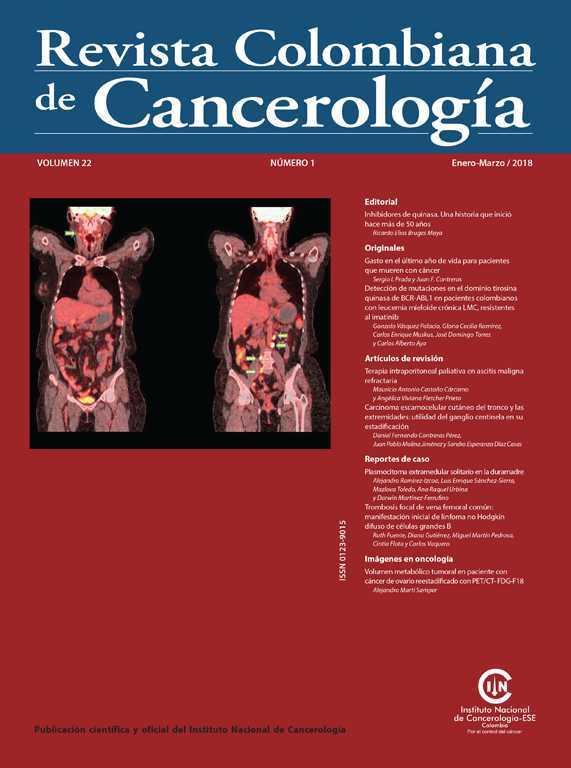
Tumour metabolic volume in a patient with ovarian cancer restaged with PET/CT- FDG-F-18
DOI:
https://doi.org/10.35509/01239015.175Keywords:
Ovarian carcinoma, Recurrence, Metastases, Positron emission tomographyAbstract
The F18-FDG PET/CT is a very useful tool in the re-staging process of cases that are suspected of relapse in the context of ovarian carcinoma. It has an excellent diagnostic and prognostic performance, providing quantitative data such as tumour metabolic volume (MTV) and total lesion glycolysis (TLG). The maximum standardised uptake value (SUVmax) is routinely reported in tumour lesions; however it must be taken into account that it is unlikely that a single pixel accurately reflects the activity of metabolically heterogeneous tumours. In order to resolve the dilemma of the use of the SUVmax, several quantitative parameters have been introduced into PET in order to estimate the measurement of the biology of the neoplasm in a more exact and objective way. In recent years, emphasis has been placed on tumour metabolic load through the use of MTV and TLG as prognostic markers and predictors of relapse.
Author Biography
Alejandro Martí Samper, Instituto Nacional de Cancerología
Servicio de Medicina Nuclear, Instituto Nacional de Cancerología, Bogotá, D.C., Colombia
References
Abdelhafez Y, Tawakol A, Osama A, Hamada E, El-Refaei S. Role of 18F-FDG PET/CT in the detection of ovarian cancer recurrence in the setting of normal tumor markers. The Egyptian Journal of Radiology and Nuclear Medicine. 2016;47:1787-94.
https://doi.org/10.1016/j.ejrnm.2016.08.013
Cho SM, Ha HK, Byun JY, Lee JM, Kim CJ, Nam-Koong SE, et al. Usefulness of FDG PET for assessment of early recurrent epithelial ovarian cancer. AJR Am J Roentgenol. 2002;179:391-5.
https://doi.org/10.2214/ajr.179.2.1790391
Rusu D, Carlier T, Colombié M, Goulon D, Fleury V, Rousseau N, et al. Clinical and Survival Impact of FDG PET in Patients with Suspicion of Recurrent Ovarian Cancer: A 6-Year Follow-Up. Front Med (Lausanne). 2015;2:46.
https://doi.org/10.3389/fmed.2015.00046
Tawakol A, Abdelhafez YG, Osama A, Hamada E, El-Refaei S. Diagnostic performance of 18F-FDG PET/contrast-enhanced CT versus contrast-enhanced CT alone for post-treatment detection of ovarian malignancy. Nucl Med Commun. 2016;37: 453-60.
https://doi.org/10.1097/MNM.0000000000000477
Fulham MJ, Carter J, Baldey A, Hicks RJ, Ramshaw JE, Gibson M. The impact of PET-CT in suspected recurrent ovarian cancer: A prospective multi-centre study as part of the Australian PET Data Collection Project. Gynecol Oncol. 2009;112:462-8.
https://doi.org/10.1016/j.ygyno.2008.08.027
Herrmann K, Benz MR, Krause BJ, Pomykala KL, Buck AK, Czernin J. (18)F-FDG-PET/CT in evaluating response to therapy in solid tumors: Where we are and where we can go. Q J Nucl Med Mol imaging. 2011;55:620-32.
Gallicchio R, Nardelli A, Venetucci A, Capacchione D, Pelagalli A, Sirignano C, et al. F-18 FDG PET/CT metabolic tumor volume predicts overall survival in patients with disseminated epithelial ovarian cancer. Eur J Radio. 2017;93:107-13.
https://doi.org/10.1016/j.ejrad.2017.05.036
Vargas HA, Burger IA, Goldman DA, Miccò M, Sosa RE, Weber W, et al. Volume-based quantitative FDG PET/CT metrics and their association with optimal debulking and progression-free survival in patients with recurrent ovarian cancer undergoing secondary cytoreductive surgery. Eur Radiol. 2015;25: 3348-53.
https://doi.org/10.1007/s00330-015-3729-9
Lee JW, Cho A, Lee JH, Yun M, Lee JD, Kim YT, et al. The role of metabolic tumor volume and total lesion glycolysis on 18F-FDG PET/CT in the prognosis of epithelial ovarian cancer. Eur J Nucl Med Mol Imaging. 2014;41:1898-906.
How to Cite
Downloads
Downloads
Published
Issue
Section
License
Todos los derechos reservados.


