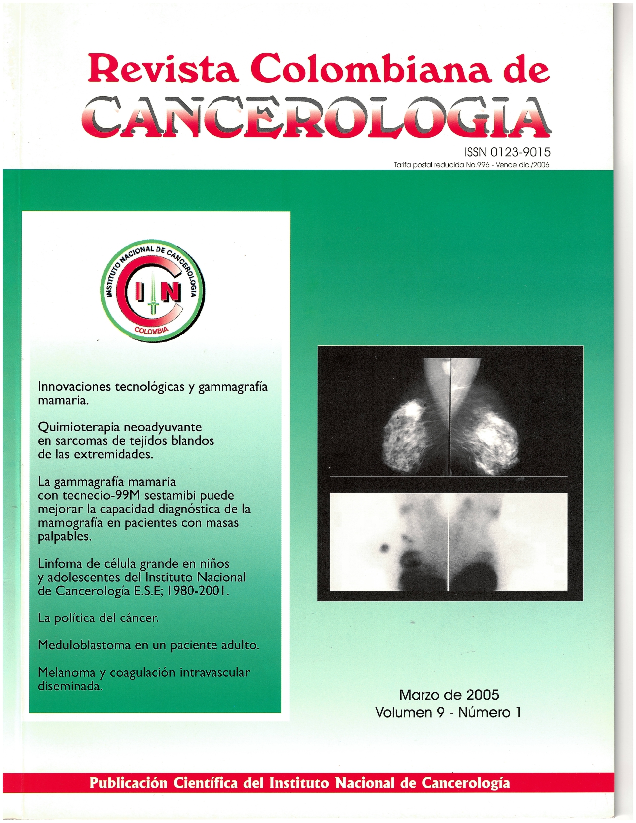
La gammagrafía mamaria con tecnecio-99m sestamibi puede mejorar la capacidad diagnóstica de la mamografía en pacientes con masas palpables
Palabras clave:
neoplasias de la mama, tecnecio Tc 99m, sestamobi, uso diagnóstico, cintigrafía, radioisótoposResumen
Objetivo: Evaluar la capacidad diagnóstica de la gammagrafía mamaria con tecnecio-99m sestamibi (99mTc-MIBI) comparativamente con la mamografía en mujeres con masas palpables que consultan al Instituto Nacional de Cancerología por sospecha clínica de cáncer de mama.
Métodos: Se evaluaron 45 pacientes consecutivos con masas palpables en los senos. A todos se les practicó una mamografía bilateral cuyos resultados se clasificaron de acuerdo con el Sistema de Información e Interpretación de Imágenes Mamarias (Bi-rads). Posteriormente, se les realizó una gammagrafía mamaria con imágenes estáticas obtenidas 10 y 90 minutos después de la aplicación intravenosa de 20 mCi de 99mTc-MIBI. El diagnóstico final se estableció mediante una biopsia percutánea con aguja cortante.
Resultados: Se demostró cáncer en 41 de las 49 lesiones estudiadas. La gammagrafía mamaria tuvo una sensibilidad del 95% y un valor predictivo positivo del 91%. La sensibilidad de la mamografía fue del 76% con un valor predictivo positivo del 100%. De los 10 resultados falsos negativos de la mamografía, 8 se presentaron en pacientes con mamas densas, nodulares o de patrón mixto: todos fueron correctamente identificados como cáncer por la gammagrafía.
Conclusión: La gammagrafía mamaria puede incluirse en el estudio de las pacientes con masas palpables, especialmente cuando tienen mamas densas, nodulares o de patrón mixto; clasificadas por la mamografía como Bi-rads 1, 2, o 3.
Biografía del autor/a
Amelia de los Reyes, Instituto Nacional de Cancerología
Instituto Nacional de Cancerología E. S. E., Grupo de Medicina Nuclear, Bogotá, D.C., Colombia.
Augusto Llamas, Instituto Nacional de Cancerología
Instituto Nacional de Cancerología E. S. E., Grupo de Medicina Nuclear, Bogotá, D.C., Colombia.
Referencias bibliográficas
Globocan 2002. Disponible en: <http://wwwdep.iarc.fr.
Olsen O, Gptzsche PC. Screening for breast cancer with mammography (Cochrane Review). In: The Cochrane Library. Issue 1. Oxford: Update Software; 2005.
Kolb TM, Lichy J, Newhouse JH. Comparison of the performance of screening mammography, physical examination and breast US, and evaluation of factors that influence them: an analysis of 27,825 patient evaluations. Radiology 2002;225:165-175.
https://doi.org/10.1148/radiol.2251011667
Verbeek ALM. The influence of breast density on the sensitivity of mammography screening. Eur J Cancer 2004;40 Supl 2:57.
https://doi.org/10.1016/S1359-6349(04)90621-0
Kopans D. The positive predictive value of mammography. AJR 1992;158:521-526.
https://doi.org/10.2214/ajr.158.3.1310825
Prats E, Aisa F, Abós MA, Villavieja L, Garcia López F, Asenjo MJ, et al. Mammography and 99mTc-MIBI scintimmamography in suspected breast cancer. J Nucl Med 1999;40:296-301.
Polan RL, Klein BD, Richman RH. Scintimammography in patients with minimal mammographic or clinical findings. Radiographics 2001;21:641-653.
https://doi.org/10.1148/radiographics.21.3.g01ma26641
Liberman M, Sampalis F, Mulder DS, Sampalis JS. Breast cancer diagnosis by scintimammography: a meta-analysis and review of the literature. Breast Cancer Res Treat 2003;80:115-126.
https://doi.org/10.1023/A:1024417331304
Sampalis FS, Denis R, Picard D, Fleiszer D, Martin G, Nassif E, et al. International prospective evaluation of scintimammography with (99m) technetium sestamibi. Am J Surg 2003;185:544-549.
https://doi.org/10.1016/S0002-9610(03)00077-1
American College of Radiology (ACR). ACR Birads-Mammography. In: Breast Imaging Reporting and Data System. Breast Imaging Atlas. 4ed. Reston (Virginia): American College of Radiology; 2003.
Lanin DR, Harns RP, Swanson FH, Edwards MS, Swanson MS, Pories WJ. Difficulties in diagnosis of carcinoma of the breast in patient less than fifty years of age. Surg Gynecol Obstet 1993; 177:457-462.
Waxman AD. The role of 99mTc-Methoxyisobutilisonitrile in imaging breast cáncer. Sem Nucí Med 1997;27:40-54.
https://doi.org/10.1016/S0001-2998(97)80035-9
Biersak HJ, Palmedo H, Bender H, Krause T. Nuclear nedicine and breast cáncer. Nucl Med Ann 2000;41:1973-1979.
Edelken S. Mammography and palpable cancer of the breast. Cancer 1988;61:263-265.
https://doi.org/10.1002/1097-0142(19880115)61:2<263::AID-CNCR2820610211>3.0.CO;2-Z
Coveney EC, Geraghty JG, O'Laoide R, Hourihane JB, O Higgins NJ. Reasons underlying negative mammography in patients with palpable breast cancer. Clin Radiol 1994;49:123-125.
https://doi.org/10.1016/S0009-9260(05)83454-3
The Steering Committee on Clinical Practice Guidelines for the Care and Treatment of Breast Cancer. Clinical practice guidelines for the care and treatment of breast cancer. CMAJ 1998; 158 Supl: 3-8.
Thurfjell E. Breast densitiy and the risk of breast cancer. N Engl J Med 2002;347:866.
https://doi.org/10.1056/NEJMp020093
Schillaci O, Buscombe JR. Breast scintigraphy today: indications and limitations. Eur J Nucl Med Mol Imaging 2004;31Supl:35-45.
https://doi.org/10.1007/s00259-004-1525-x
Rhodes DJ, O'ConnorMK, Phillips SW, SmithRL, Collins DA. Molecular breast imaging: a new technique using technetium Tc-99m scintimammography to detect small tumors of the breast. Mayo Clin Proc 2005;80:24-30.
https://doi.org/10.1016/S0025-6196(11)62953-4
Boyd NF, Dite GS, Stone J, Gunasekara A, English DR, McCredie MR, et al. Heritability of mammographic density, a risk factor for breast cancer. N Engl J Med 2002;347:886-894.
https://doi.org/10.1056/NEJMoa013390
Khalkhali I, Villanueva-Meyer J, Edell SL, Conolly JL, Schnitt S J, Baum JK, et al. Diagnostic accuracy of 99mTc-sestamibi breast imaging: multicentric trial results. J Nucl Med 2000;41:1973-1979.
Primic-Zakelj M. Screening mammography to early detection of breast cancer. Ann Oncol 1999; 10 Supl: 121-127.
https://doi.org/10.1023/A:1008310503380
Sun SS, Hsieh JF, Tsai SC, Ho YJ, Kao CH. Expression of drug resistance protein related to 99mTc-MIBI breast imaging. Anticancer Res 2000;20:2021-2026.
Cómo citar
Descargas
Descargas
Publicado
Número
Sección
Licencia
Todos los derechos reservados.


