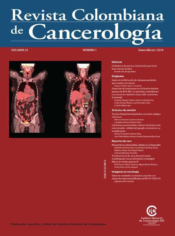
Volumen metabólico tumoral en paciente con cáncer de ovario reestadificado con PET/CT- FDG-F18
DOI:
https://doi.org/10.35509/01239015.175Palabras clave:
Carcinoma de ovario, Recurrencia, Metástasis, Tomografía por emisión de positronesResumen
El F18-FDG PET/CT es una herramienta de gran utilidad en el proceso de reestadificación de los casos que tienen sospecha de recaída en el contexto de carcinoma de ovario con excelente rendimiento diagnóstico y pronóstico, suministrando datos cuantitativos como el volumen metabólico tumoral (MTV) y lesión glicolitica total (TLG). De manera rutinaria se reporta el valor de captación estandarizado máximo (SUVmax) en las lesiones tumorales, sin embargo, hay que tener en cuenta que es improbable que un solo píxel refleje la actividad de tumores metabólicamente heterogéneos con precisión. Con el fin de resolver el dilema del uso del SUVmax, se han introducido varios parámetros cuantitativos en PET para estimar la medición de manera más exacta y objetiva de la biología de la neoplasia. En años recientes se ha hecho énfasis en la carga metabólica tumoral mediante el uso del MTV y la TLG como marcadores pronósticos y predictores de recaída.
Biografía del autor/a
Alejandro Martí Samper, Instituto Nacional de Cancerología
Servicio de Medicina Nuclear, Instituto Nacional de Cancerología, Bogotá, D.C., Colombia
Referencias bibliográficas
Abdelhafez Y, Tawakol A, Osama A, Hamada E, El-Refaei S. Role of 18F-FDG PET/CT in the detection of ovarian cancer recurrence in the setting of normal tumor markers. The Egyptian Journal of Radiology and Nuclear Medicine. 2016;47:1787-94.
https://doi.org/10.1016/j.ejrnm.2016.08.013
Cho SM, Ha HK, Byun JY, Lee JM, Kim CJ, Nam-Koong SE, et al. Usefulness of FDG PET for assessment of early recurrent epithelial ovarian cancer. AJR Am J Roentgenol. 2002;179:391-5.
https://doi.org/10.2214/ajr.179.2.1790391
Rusu D, Carlier T, Colombié M, Goulon D, Fleury V, Rousseau N, et al. Clinical and Survival Impact of FDG PET in Patients with Suspicion of Recurrent Ovarian Cancer: A 6-Year Follow-Up. Front Med (Lausanne). 2015;2:46.
https://doi.org/10.3389/fmed.2015.00046
Tawakol A, Abdelhafez YG, Osama A, Hamada E, El-Refaei S. Diagnostic performance of 18F-FDG PET/contrast-enhanced CT versus contrast-enhanced CT alone for post-treatment detection of ovarian malignancy. Nucl Med Commun. 2016;37: 453-60.
https://doi.org/10.1097/MNM.0000000000000477
Fulham MJ, Carter J, Baldey A, Hicks RJ, Ramshaw JE, Gibson M. The impact of PET-CT in suspected recurrent ovarian cancer: A prospective multi-centre study as part of the Australian PET Data Collection Project. Gynecol Oncol. 2009;112:462-8.
https://doi.org/10.1016/j.ygyno.2008.08.027
Herrmann K, Benz MR, Krause BJ, Pomykala KL, Buck AK, Czernin J. (18)F-FDG-PET/CT in evaluating response to therapy in solid tumors: Where we are and where we can go. Q J Nucl Med Mol imaging. 2011;55:620-32.
Gallicchio R, Nardelli A, Venetucci A, Capacchione D, Pelagalli A, Sirignano C, et al. F-18 FDG PET/CT metabolic tumor volume predicts overall survival in patients with disseminated epithelial ovarian cancer. Eur J Radio. 2017;93:107-13.
https://doi.org/10.1016/j.ejrad.2017.05.036
Vargas HA, Burger IA, Goldman DA, Miccò M, Sosa RE, Weber W, et al. Volume-based quantitative FDG PET/CT metrics and their association with optimal debulking and progression-free survival in patients with recurrent ovarian cancer undergoing secondary cytoreductive surgery. Eur Radiol. 2015;25: 3348-53.
https://doi.org/10.1007/s00330-015-3729-9
Lee JW, Cho A, Lee JH, Yun M, Lee JD, Kim YT, et al. The role of metabolic tumor volume and total lesion glycolysis on 18F-FDG PET/CT in the prognosis of epithelial ovarian cancer. Eur J Nucl Med Mol Imaging. 2014;41:1898-906.
Cómo citar
Descargas
Publicado
Número
Sección
Licencia
Todos los derechos reservados.

| Estadísticas de artículo | |
|---|---|
| Vistas de resúmenes | |
| Vistas de PDF | |
| Descargas de PDF | |
| Vistas de HTML | |
| Otras vistas | |


