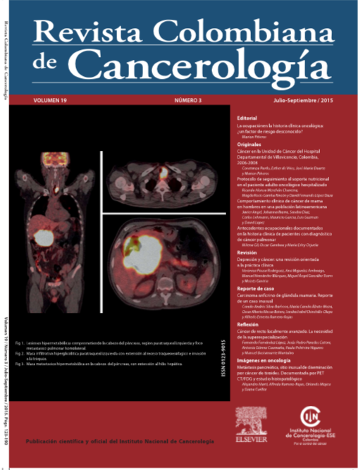
Metástasis pancreática, sitio inusual de diseminación por cáncer de tiroides. Documentada por PET CT/FDG y estudio histopatológico
Palabras clave:
Carcinoma papilar de tiroides, Metástasis, Tomografía por emisión de positronesResumen
El cáncer diferenciado de tiroides (CDT) es la neoplasia endocrina maligna encontrada con mayor frecuencia, generalmente tiene un comportamiento lento en su evolución permitiendo realizar manejo quirúrgico y terapia ablativa con Iodo 131, lográndose la remisión completa en la gran mayoría de los casos. Un pequeño porcentaje de estas neoplasias presenta un comportamiento agresivo al registrar metástasis a distancia con localización principalmente en pulmón, hueso y cerebro y con focos de desdiferenciación celular, lo cual empobrece su pronóstico y limita las opciones terapéuticas en este tipo de tumores. En el proceso de seguimiento del CDT, la tomografía por emisión de positrones con análogo de glucosa (PET/CT con F18-FDG) se ha constituido en una herramienta diagnóstica y pronóstica de imagen eficaz. Presentamos el caso de un paciente masculino de 50 años de edad con cáncer papilar de tiroides y enfermedad metastásica en páncreas, sitio inusual de diseminación para esta patología.
Biografía del autor/a
Alejandro Martí, Instituto Nacional de Cancerología
Servicio de Medicina Nuclear, Instituto Nacional de Cancerología, Bogotá D. C., Colombia
Alfredo Romero Rojas, Instituto Nacional de Cancerología
Servicio de Patología, Instituto Nacional de Cancerología, Bogotá D. C., Colombia
Orlando Mojica, Fundación Universitaria Sanitas
Medicina Nuclear, Fundación Universitaria Sanitas, Bogotá D. C., Colombia
Diana Cuéllar, Instituto Nacional de Cancerología
Servicio de Medicina Nuclear, Instituto Nacional de Cancerología, Bogotá D. C., Colombia
Referencias bibliográficas
Mazzaferri EL, Kloos RT. Clinical review 128: Current approaches to primary therapy for papillary and follicular thyroid cancer. J Clin Endocrinol Metab. 2001;86(4):1447-63.
https://doi.org/10.1210/jcem.86.4.7407
Globocan 2012 - Home Internet. consulta el 20 de enero de 2014. Disponible en: http://globocan.iarc.fr/Default.aspx
Murillo Moreno RH, Piñeros Petersen M, Acosta Peñaloza JA, Castellanos Herrera VH. Anuario estadístico 2010. Bogotá, D.C., Colombia: Instituto Nacional de Cancerología, Ministerio de Salud y Protección Social; 2010. p. 96.
LiVolsi VA. Papillary thyroid carcinoma: an update. Mod Pathol. 2011;24 Suppl 2:S1-9.
https://doi.org/10.1038/modpathol.2010.129
Rahman GA, Abdulkadir AY, Olatoke SA, Yusuf IF, Braimoh KT. Unusual cutaneous metastatic follicular thyroid carcinoma. J Surg Tech Case Rep. 2010;2(1):35-8.
https://doi.org/10.4103/2006-8808.63724
Benbassat CA, Mechlis-Frish S, Hirsch D. Clinicopathological characteristics and long-term outcome in patients with distant metastases from differentiated thyroid cancer. World J Surg. 2006;30(6):1088-95.
https://doi.org/10.1007/s00268-005-0472-4
Muresan MM, Olivier P, Leclère J, Sirveaux F, Brunaud L, Klein M, et al. Bone metastases from differentiated thyroid carcinoma. Endocr Relat Cancer Internet. 2008;15(1):37-49.
https://doi.org/10.1677/ERC-07-0229
Durante C, Haddy N, Baudin E, Leboulleux S, Hartl D, Travagli JP, et al. Long-term outcome of 444 patients with distant metastases from papillary and follicular thyroid carcinoma: benefits and limits of radioiodine therapy. J Clin Endocrinol Metab. 2006;91(8):2892-9.
https://doi.org/10.1210/jc.2005-2838
Lopez-Penabad L, Chiu AC, Hoff AO, Schultz P, Gaztambide S, Ordoñez NG, et al. Prognostic factors in patients with Hürthle cell neoplasms of the thyroid. Cancer. 2003;97(5):1186-94.
https://doi.org/10.1002/cncr.11176
Tunio MA, Alasiri M, Riaz K, Alshakweer W. Pancreas as delayed site of metastasis from papillary thyroid carcinoma. Case Rep Gastrointest Med. 2013 Jan;2013:386263.
https://doi.org/10.1155/2013/386263
Borschitz T, Eichhorn W, Fottner C, Hansen T, Schad A, Schadmand-Fischer S, et al. Diagnosis and treatment of pancreatic metastases of a papillary thyroid carcinoma. Thyroid. 2010;20(1):93-8.
https://doi.org/10.1089/thy.2009.0026
Siddiqui AA, Olansky L, Sawh RN, Tierney WM. Pancreatic metastasis of tall cell variant of papillary thyroid carcinoma: diagnosis by endoscopic ultrasound-guided fine needle aspiration. JOP. 2006;7(4):417-22.
Chen L, Brainard JA. Pancreatic metastasis from papillary thyroid carcinoma diagnosed by endoscopic ultrasound-guided fine needle aspiration: a case report. Acta Cytol. 2010;54(4):640-4.
https://doi.org/10.1159/000325192
Adsay NV, Andea A, Basturk O, Kilinc N, Nassar H, Cheng JD. Secondary tumors of the pancreas: an analysis of a surgical and autopsy database and review of the literature. Virchows Arch. 2004;444(6):527-35.
https://doi.org/10.1007/s00428-004-0987-3
Marcus C, Whitworth PW, Surasi DS, Pai SI, Subramaniam RM. PET/CT in the management of thyroid cancers. AJR Am J Roentgenol. 2014;202(6):1316-29.
https://doi.org/10.2214/AJR.13.11673
Abraham T, Schöder H. Thyroid cancer-indications and opportunities for positron emission tomography/computed tomography imaging. Semin Nucl Med Elsevier Inc. 2011;41(2):121-38.
https://doi.org/10.1053/j.semnuclmed.2010.10.006
Edward J. Escott Role of positron emission tomography/ computed tomography (PET/CT) in head and neck cancer. Radiol Clin North Am. 2013;51(5):881-93.
https://doi.org/10.1016/j.rcl.2013.05.002
Robbins RJ, Wan Q, Grewal RK, Reibke R, Gonen M, Strauss HW, et al. Real-time prognosis for metastatic thyroid carcinoma based on 2-18 Ffluoro-2-deoxy-D-glucose-positron emission tomography scanning. J Clin Endocrinol Metab. 2006;91(2):498-505.
https://doi.org/10.1210/jc.2005-1534
Grünwald F, Menzel C, Bender H, Palmedo H, Willkomm P, Ruhlmann J, et al. Comparison of 18 FDG-PET with 131 iodine and 99 mTc-sestamibi scintigraphy in differentiated thyroid cancer. Thyroid. 1997;7(3):327-35.
https://doi.org/10.1089/thy.1997.7.327
Feine U, Lietzenmayer R, Hanke JP, Wöhrle H, Müller Schauenburg W. 18 FDG whole-body PET in differentiated thyroid carcinoma Flipflop in uptake patterns of 18 FDG and 131 I. Nuklearmedizin. 1995;34(4):127-34.
https://doi.org/10.1055/s-0038-1629813
Wang W, Macapinlac H, Larson SM, Yeh SD, Akhurst T, Finn RD, et al. 18 F- 2-fluoro-2-deoxy-D-glucose positron emission tomography localizes residual thyroid cancer in patients with negative diagnostic 131 I whole body scans and elevated serum thyroglobulin levels. J Clin Endocrinol Metab. 1999;84(7):2291-302.
Cooper DS, Doherty GM, Haugen BR, Kloos RT, Lee SL, Mandel SJ, et al. Revised American Thyroid Association management guidelines for patients with thyroid nodules and differentiated thyroid cancer. Thyroid. 2009;19(11):1167-214.
https://doi.org/10.1089/thy.2009.0110
Giovanella L, Trimboli P, Verburg FA, Treglia G, Piccardo A, Foppiani L, et al. Thyroglobulin levels and thyroglobulin doubling time independently predict a positive 18F-FDG PET/CT scan in patients with biochemical recurrence of differentiated thyroid carcinoma. Eur J Nucl Med Mol Imaging. 2013;40(6):874-80.
https://doi.org/10.1007/s00259-013-2370-6
Vural GU, Akkas BE, Ercakmak N, Basu S, Alavi A. Prognostic significance of FDG PET/CT on the follow-up of patients of differentiated thyroid carcinoma with negative 131I whole-body scan and elevated thyroglobulin levels: correlation with clinical and histopathologic characteristics and long-term follow-up data. Clin Nucl Med. 2012;37:953-9.
https://doi.org/10.1097/RLU.0b013e31825b2057
Schönberger J, Rüschoff J, Grimm D, Marienhagen J, Rümmele P, Meyringer R, et al. Glucose transporter 1 gene expression is related to thyroid neoplasms with an unfavorable prognosis: an immunohistochemical study. Thyroid. 2002;12(9):747-54.
https://doi.org/10.1089/105072502760339307
Urhan M, Basu S, Alavi A. PET Scan in Thyroid Cancer. PET Clin. 2012;7:453-61.
Cómo citar
Descargas
Descargas
Publicado
Número
Sección
Licencia
Todos los derechos reservados.


